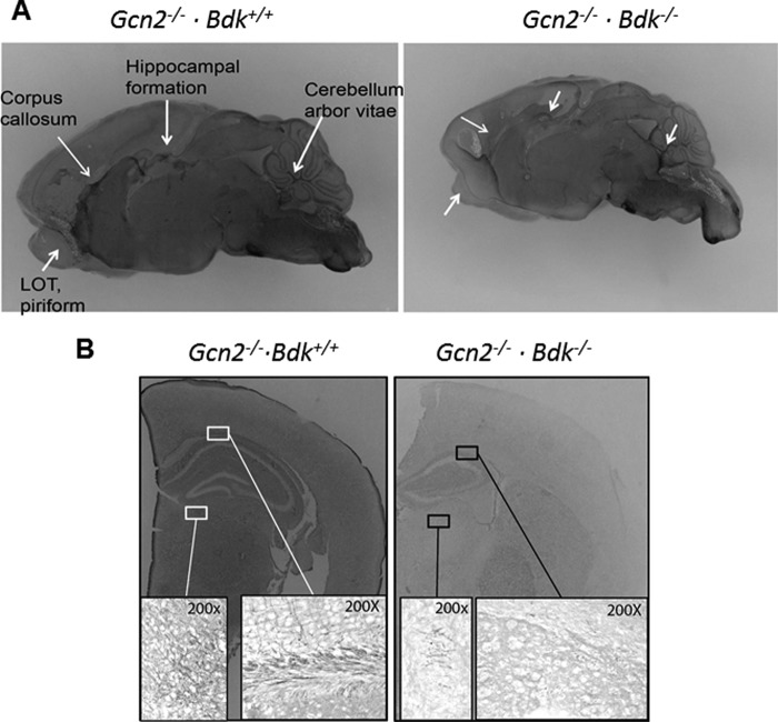
FIGURE 4.
GBDK mice demonstrate hypomyelination in white matter regions of the brain. A, representative gold chloride staining of bisected sagittal hemispheres of brains imaged using a dissecting microscope set to ×1 magnification. B, representative coronal sections taken rostral to bregma using a dissecting microscope set to ×2 magnification. Higher magnification (×200) under a light microscope revealed the presence of myelin fibers in the corpus callosum and hippocampal formation. Images are representative of three mice per genotype at postnatal days 11–14.

