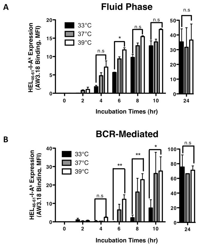
Figure 8. Fever Range Temperature Accelerates the Kinetics of BCR-mediated Antigen Presentation.
Splenocytes from B10.Br (Panel A) or MD4.B10.Br (Panel B) mice were pre-incubated for 2 hours at the indicated temperature. Cells were then pulsed with antigen [MD4.B10.Br – 100 nM HEL for BCR-mediated processing, B10.Br – 100 μM HEL + 10 μg/ml anti-BCR for fluid phase processing and BCR signaling, respectively (12)] for the indicated time at the indicated temperature. Cells were harvested and the level of HEL46–61–I-Ak complex expression was determined by staining with the HEL46–61–I-Ak complex-specific mAb Aw3.18 (15) and analysis of Aw3.18 binding by flow cytometry (12). Shown is the average MFI of Aw3.18 staining of B220+ B cells +/− S.E.M. for 3 independent experiments. The level of Aw3.18 binding detected with the 33°C and 39°C samples at each time point/antigen dose was compared by a Student’s t-test (** = p value of <0.01, *= p value of <0.05, n.s = not significant with a p value of >0.05). The total level of I-Ak class II expression was also monitored and did not vary by more than +/− 10% between samples.

