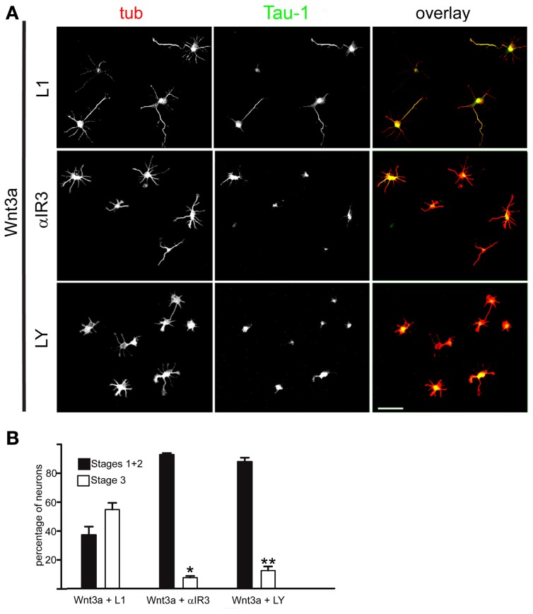
Figure 3.
An IGF-1r blocking antibody or the PI3k inhibitor LY294002 hindered the polarizing effects of Wnt3a. (A) Double immunofluorescence micrographs of hippocampal neurons after 20 h in culture showing the distribution of the neuronal marker β-III tubulin (tub) and the axonal marker Tau-1, Cells were cultured in medium containing 1.35 nM Wnt3a plus an antibody to L1 (top), 1.35 nM Wnt3a plus the IGF-1r blocking antibody αIR3 (middle), or 1.35 nM Wnt3a plus the PI3k inhibitor LY294002 (bottom). (B) Percentage (±sem) of neurons at different stages of differentiation grown for 20 h in medium containing 1.35 nM Wnt3a plus the anti-L1 antibody, 1.35 nM Wnt3a plus the IGF-1r blocking antibody αIR3, or 1.35 nM Wnt3a plus the PI3k inhibitor LY294002 (n = 3 independent experiments). At least 100 cells were scored for each condition. *p < 0.001 compared with Wnt3a + L1; **p < 0.001 compared with Wnt3a (Figure 1B, right). Scale bar: 100 μm.

