Figure 7.

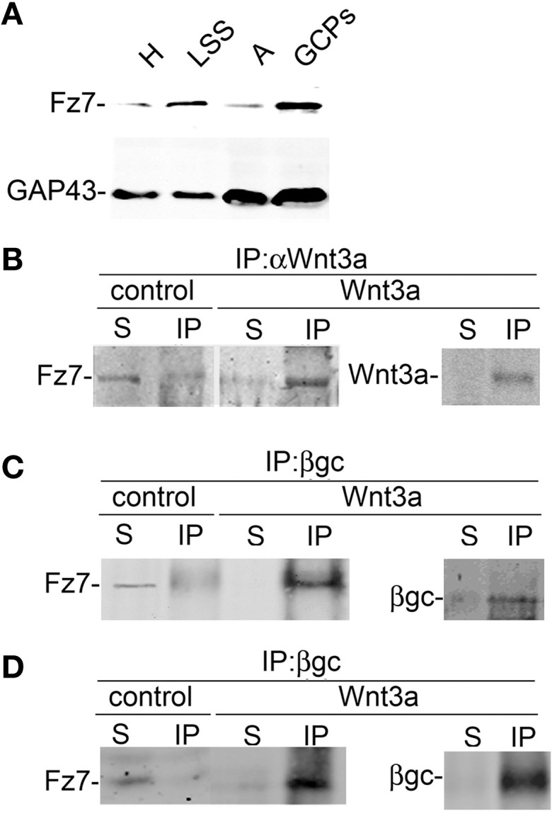
Activation with Wnt3a triggers the formation of a IFG-1r/Wnt3a/Fz7 complex. (A) Fz7 is enriched in GCPs. Western blot analysis of the distribution of the Wnt receptor Fz7 in different subcellular fractions of fetal brain: homogenate (H), low-speed supernatant (LSS), A-fraction (A), and growth cone particles (GCPs). Equal amounts of protein were loaded in all lines. Note the enrichment of Fz7 in GCPs around fourfold compared to (H). A blot showing the distribution of GAP-43, a protein highly enriched in GCPs, was included for comparison. (B,C) Wnt3a triggered the formation of a complex containing IGF-1r-Wnt3a-Fz7. Growth cone particles were incubated in control buffer or in the presence of 1.35 nM Wnt3a and immunoprecipitated with (B) Anti Wnt3a and probed with an antibody to the Wnt receptor Fz7 and (C) immunoprecipitated with β gc antibody to pull down IGF-1r and probed with anti Fz7. Immunoprecipitation of Wnt3a and β gc are shown to confirm successful IP and as a loading control. IP, immunoprecipitate; S, soluble fraction after immunoprecipitation. (D) Total protein from hippocampal neurons cultured for 20 h in the presence of 1.35 nm Wnt3a, deprived of any growth factor for 4 h, and stimulated (or not) with 1.35 nM Wnt3a for 5 min immunoprecipitated with anti β gc antibody to pull down the IGF-1r and probed with anti Fz7. Immunoprecipitation of β gc is shown to confirm successful IP and as a loading control. IP, immunoprecipitate; S, soluble fraction after immunoprecipitation. All blots are representative of at least three independent experiments.
