
Figure 5:
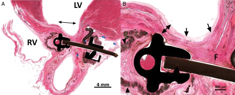
Microscopic tissue examination of the implant sites (H&E Stain, 10x). (A) There is reduction of the infracted area and fusion of the apposed infarct margins (double arrow). (B) There is healing of the fused infarct wall with fibrous tissue healing (F) and coverage of the endocardium by a small amount of neointima (arrows). There is moderate compressive attenuation of the ventricular wall (double arrow). Inflammation around the velour cover is minimal (arrowheads).