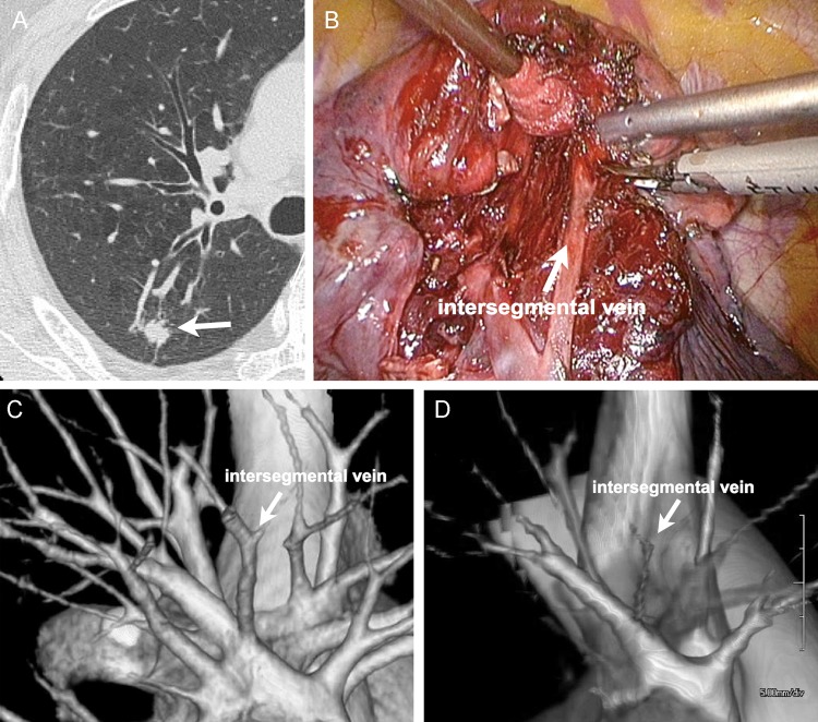
Figure 1:
S.2 segmentectomy of the right lobe of a 56-year old male. (A) CT image shows a 12-mm primary lung tumour (adenocarcinoma) located in S.2. (B) Interoperative view. The intersegmental plane was dissected while preserving the intersegmental vein between S.2 and S.3. (C) 3D vasculature image from area detector CT (ADCT) data clearly shows the intersegmental vein between two segments. This image was scored as 5. (D) 3D vasculature image from multidetector CT (MDCT) data clearly shows the intersegmental vein between two segments. This image was scored as 3.

