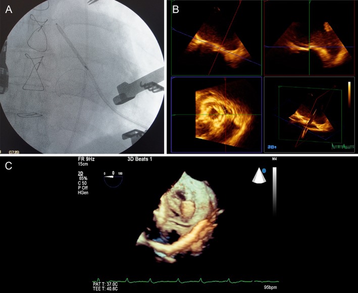
Figure 1:
(A) Fluoroscopic view of the guidewire crossing the paravalvular defect. (B) Transoesophageal tridimensional echocardiography, showing defect identification. (C) Transoesophageal tridimensional echocardiography, showing the guidewire crossing the paravalvular defect and initial deployment of the Amplatzer Vascular Plug II.

