
Figure 1:
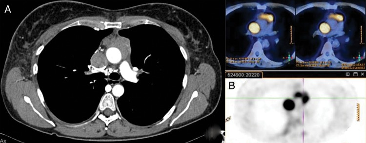
(A) Chest CT scan shows superior vena cava (SVC) thrombosis until the right atrium and a separate anterior mediastinal mass. (B) PET/CT scan shows two different pathological uptake values in the thymic region anteriorly to the ascendant aorta and in the SVC.