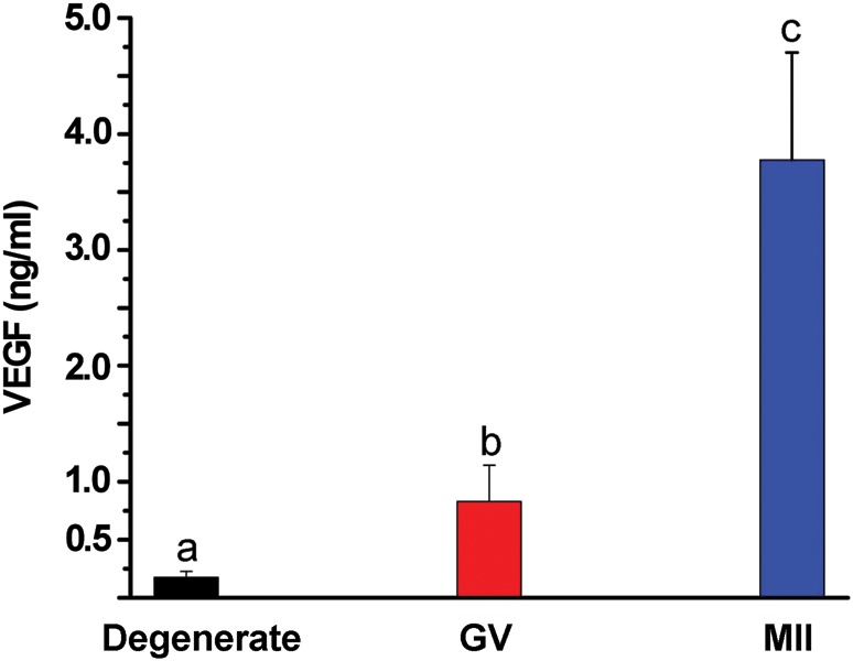
Figure 3.

VEGF-A concentrations at Week 5 from cultures of growing (slow- or fast-grow) follicles as a function of oocyte quality/maturation following exposure to 100 ng/ml hCG. Values are the mean ± SEM, n = 45, 19 and 3 for follicles from 9 monkeys providing degenerate (black), germinal-vesicle (GV, red) or metaphase II (MII, blue) oocytes, respectively, at retrieval. Significant differences between groups are denoted by different letters.
