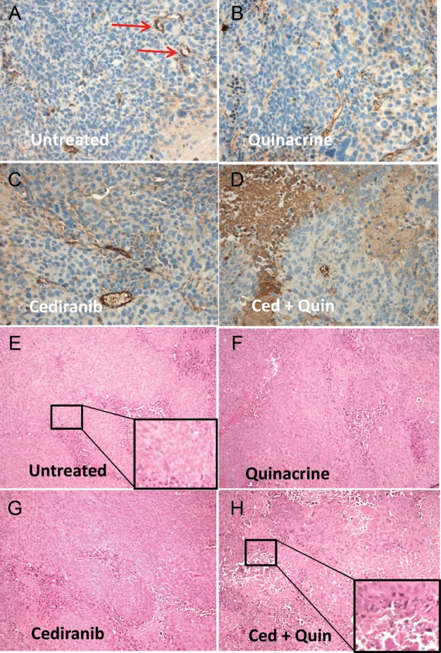
Fig. 5.

(A–D) Representative histological tumor sections with CD31 vascular staining (brown) and hematoxylin nuclear counterstain (blue) from the 4 treatment groups. Discrete staining is associated with vascular endothelial cells, whereas more diffuse and variable staining is nonspecific and associated with tumor necrosis. Microvessel examples are marked by arrows. (E–H) Representative H&E stained sections from the 4 treatment groups, indicating areas of necrosis, which appear as condensed cells in areas of vacuolated tissue. Insets indicate enlarged areas of the section as indicated.
