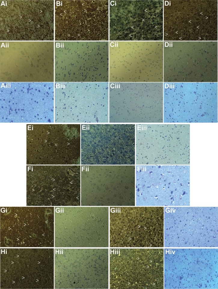
Fig. 3.
VEGF, HIF-1α, GLUT-1, SLIT3, spondin1, and Plexin-B2 levels are elevated in high-grade gliomas compared with low-grade gliomas. (A) Representative IHC staining for VEGF in a GBM (Ai), an oligodendroglioma (Aii), and normal brain (Aiii). (B) Representative IHC staining for HIF-1α in a GBM (Bi), a low-grade astrocytoma (LGA) (Bii), and normal brain (Biii). (C) Representative IHC staining for GLUT-1 in a GBM (Ci), an oligodendroglioma (Cii), and normal brain (Ciii). (D) Representative IHC staining for SLIT3 in a GBM (Di), an LGA (Dii), and normal brain (Diii). (E) Representative IHC staining for spondin1 in a GBM (Ei), an LGA (Eii), and normal brain (Eiii). (F) Representative IHC staining for Plexin-B2 in a GBM (Fi), an oligodendroglioma (Fii), and normal brain (Fiii). (G) Representative IHC staining for fibulin-1 in a GBM (Gi), an LGA (Gii, an example of low expression), an oligodendroglioma (Giii, an example of high expression), and normal brain (Giv). (H) Representative IHC staining for LINGO in a GBM (Hi), 2 oligodendrogliomas (Hii, an example of low expression; and Hiii, an example of high expression), and normal brain (Hiv). White arrow heads depict regions that highly stain for each biomarker. Magnification of all panels is 40×.

