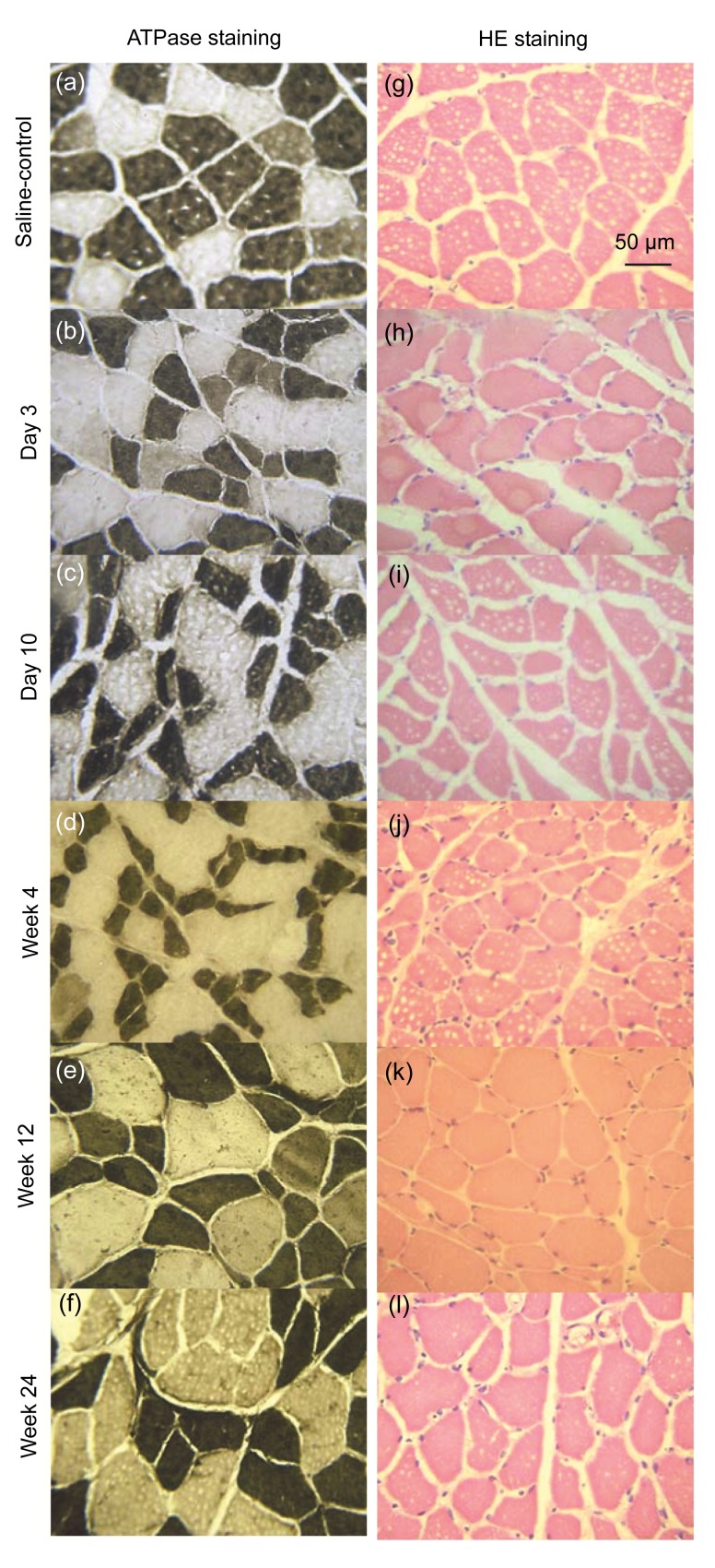
Fig. 3.
ATPase enzyme and HE stainings of gastrocnemius muscles (specimen incubated at pH 10.4)
(a) ATPase enzyme staining of the uninjected gastrocnemius muscle in the control group; (b–f) ATPase enzyme staining of the CBTX-A-injected gastrocnemius muscle in the treatment group (after 3 d, 10 d, and 4 weeks, 12 weeks, and 24 weeks, respectively); (g) HE staining of the normal saline-injected gastrocnemius muscle in the control group; (h–l) HE staining of the CBTX-A-injected gastrocnemius muscle in the treatment group (after 3 d, 10 d, and 4 weeks, 12 weeks, and 24 weeks, respectively). There were six rats in each group

