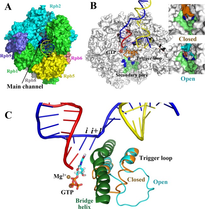
Figure 1.
Multisubunit RNAP (S. cerevisiae RNAP II). (A) Complex subunit structure and main enzyme channel. (B) Cutaway image (parts of Rpb1 and Rpb2 are missing) to show the transcription bubble, secondary pore (lime green; blue indicates basic residues important in PPi release),21 and buried active site. RNA is red, template DNA is blue, nontemplate DNA is yellow, the closed trigger loop conformation is orange, and the open trigger loop conformation is cyan. Images to the right indicate that a TEC with a closed trigger loop (orange) mostly closes the pore, and a TEC with an open trigger loop (cyan) has a more open pore with a diameter comparable to a diffusing GTP substrate. (C) RNAP active site with closed and open trigger loop conformations overlaid. Colors are as in panel B. The bridge helix is dark green. PDB structures 2E2H and 2E2J (with the open trigger loop modeled) and a PDB file from Jens Michaelis showing the intact bubble27b were used to make the images, by use of the program Visual Molecular Dynamics.94.

