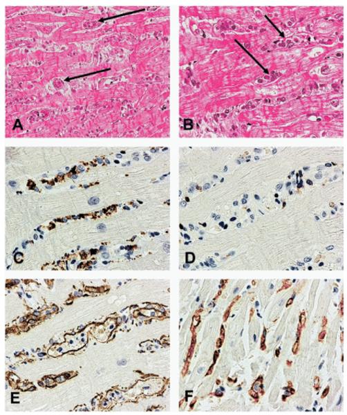
Figure 1.
Endomyocardial biopsy findings in antibody-mediated rejection. (A) Prominent intravascular cells are present in the myocardium in the absence of lymphoid cellular infiltrate (hematoxylin and eosin [H&E]; original magnification ×200). (B) Magnification of panel A at ×400 (H&E). (C) Prominence of macrophages amongst the cells with a CD68 immunoperoxidase stain (original magnification ×400). (D) Presence of only rare T lymphocytes with CD3 immunoperoxidase stain (original magnification ×400); (E) Highlighting of endothelial cells and confirmation of the macrophages’ intravascular location with the CD34 immunoperoxidase stain (original magnification ×400). (F) Deposition of C4d complement component in capillaries with the C4d immunoperoxidase stain (original magnification ×400). From: Fishbein and Kobashigawa: Current Opin. Cardiol 2004;19:166. Reprinted with permission of Wolters Kluwer.

