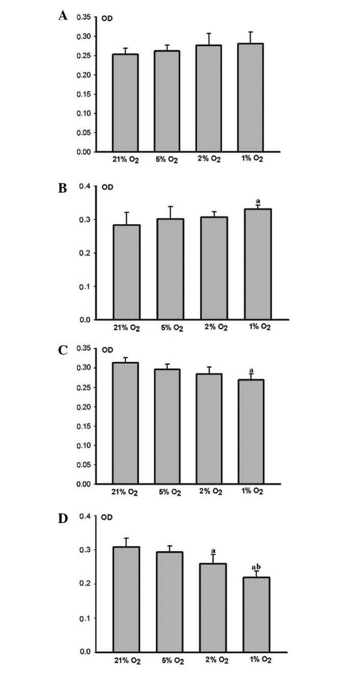
Figure 3.

Effects of different hypoxic conditions on the proliferation of human periodontal ligament fibroblasts (HPLFs) at various times. n=4, avs. 21% O2 P<0.05, bvs. 2% O2 P<0.05 (ANOVA and paired t-test). (A) At 12 h post-cultivation, the HPLFs grew more rapidly as the degree of hypoxia was increased, and there was no clear difference between the groups. (B) At 24 h post-cultivation. The HPLFs grew more rapidly as the degree of hypoxia was increased. Cell proliferation in the severe hypoxia (1% O2) group was significant. (C) At 48 h post-cultivation, the growth of HPLFs was restrained as the degree of hypoxia was increased. Cell proliferation in the severe hypoxia group was markedly restrained. (D) At 72 h post cultivation. HPLF growth was restrained as the degree of hypoxia was increased, cell proliferation in the middle (2% O2) and severe hypoxia groups was markedly restrained. The restraint was more visible in severe hypoxia.
