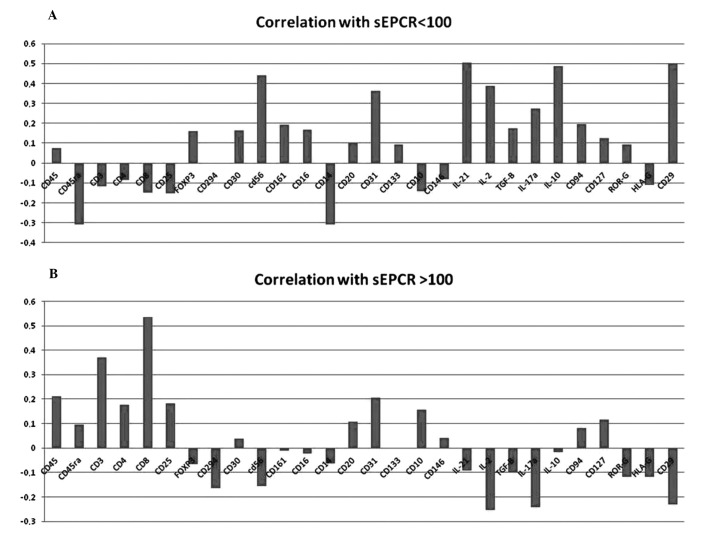
Figure 3.
Influence of plasma sEPCR in blood immune cells. Mononuclear cells from 33 patients were separated by Ficoll gradient centrifugation and then stored in 200 μ l of DMSO/FCS 50% at −80°C. Phenotyping of immuno-inflammatory cells were performed by flow cytometry the using 30 antibodies as given above. (A) When sEPCR is <100 ng/ml (group 1), plasmatic sEPCR correlated with low intensity of some cell associated markers such as CD45ra, CD3, CD8 and high intensity of with CD56 (NK cells) and cell associated IL-2, IL-10, IL-17a, IL-21 and CD29 markers (r ≤0.40). When sEPCR is >100 ng/ml (group 2), plasmatic sEPCR correlated with high intensity of CD45ra, CD3, CD8, CD25 and low intensity of CD56, IL-2, IL-10, IL-21 and CD29 markers (r ≥0.60). The expression of some cell markers such as CD4, FoxP3, CD20 and CD10 was independent of plasma sEPCR variation (r ≤0.3). A decrease in IL-17a in group-2 (B) was associated with the diminition of CD161 and ROR-γ associated cells. These results indicate that increase of plasma sEPCR was associated with a decrease of effector immune cells in blood circulation.

