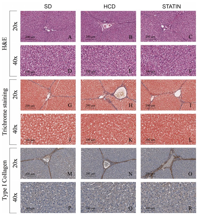
Figure 4. Hypercholesterolemia did not induce liver steatosis and fibrosis in HCD-fed pigs.
Hematoxylin and Eosin staining (A-F), Masson’s trichrome staining (G-L) and immunohistochemical analysis for type I collagen (M-R) of livers from HCD-fed pigs showed neither signs of steatosis (B, E) nor fibrosis (H, K, N, Q) having a parenchymal structure and ECM deposition comparable with those found in SD-fed (A, D, G, J, M, P) and atorvastatin-treated pigs (C, F, I, L, O, R). *P<0.05.

