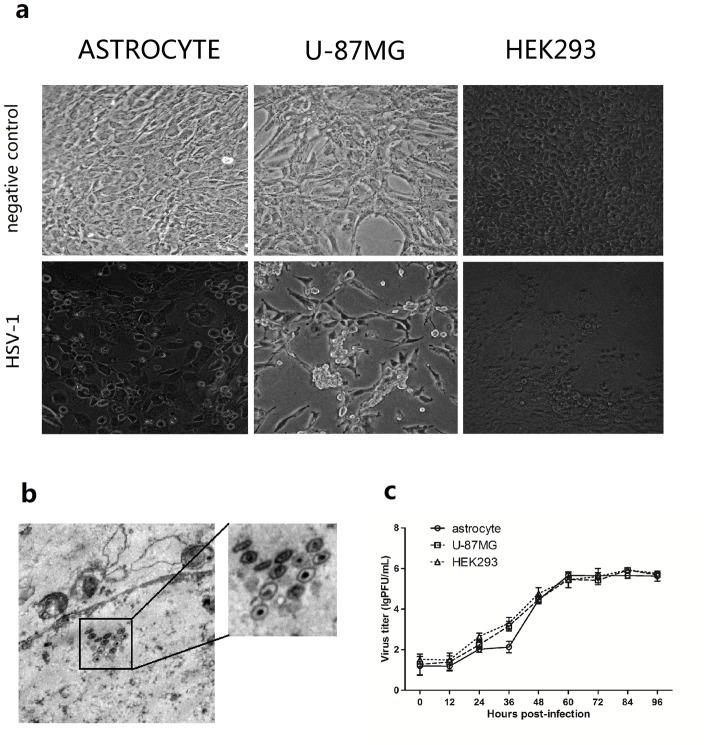
Figure 1. Replication of HSV-1 in Macaca mulatta astrocytes.
(a) HSV-1 infection of primary astrocytes. Cellular morphology of astrocyte, U-87MG and HEK293 before and after HSV-1 infection under the light microscope; Upper panels: negative control; Lower panels: 12 hours post HSV-1 infection (MOI = 1). The magnification is 200 X. (b) Virus morphology in astrocytes infected with HSV-1. The monolayer of astrocytes was infected with HSV-1 (MOI = 2). The infected astrocytes were observed at 24 h post infection using Transmission Electron Microscope (TEM), and the typical structure was observed for HSV-1 virus capsids. The magnification is 6000 X. (c) Viral replication curve in rhesus primary astrocytes, U-87MG and HEK293 cells infected with HSV-1. The primary astrocytes, U-87MG and HEK293 cells were infected with HSV-1 (MOI = 0.01). Samples were collected at 0, 12, 24, 36, 48, 60, 72, 84 and 96 hr post infection. A plaque assay was used to determine the virus titer in all of the samples to generate the viral replication curve. Error bars represent the standard deviation from triplicate samples.

