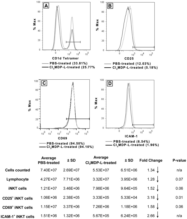
Figure 2. Kupffer cells induce the activation and accumulation of iNKT cell in biliary obstructed livers.
Animals were treated with Cl2MDP-L to deplete Kupffer cells prior to BDL; control mice received PBS. At 18 hours post-BDL, the NPCs were teased through screens, counted, purified on Percoll gradients and stained. Hepatic, CD1d-tetramer+ iNKT cells were quantified (A), and the expression of CD25 (B), CD69 (C), and ICAM-1 (D) was determined by flow cytometry. Values in parentheses denote percentages of expression. A summary of experimental results is presented in table format containing cell numbers and statistical analyses. Data are derived from three independent experiments, n = 3-6 mice/group. n/a = not available; small sample size precludes statistical analysis.

