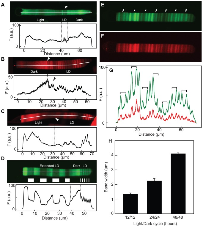
Figure 4. Effect of varying light-dark cycle on periodic axial variation.
(A) Transgenic animals expressing Rho-eGFP were housed for 2 weeks in constant darkness, switched to 24 h (12D:12L) for 3 days (LD) and then in constant light for 2 weeks prior to imaging. The intensity profile of the cell is shown below. While axial banding with spatial period 1-1.4 µm are seen in the 24 h light cycle (arrow), little variation with this spatial period was observed in constant dark or light. Note that the rate of membrane addition to the OS is higher in the light than in either cycling or constant dark conditions. (B) Transgenic animals expressing Rho-mCherry were housed in 24 h (12D:12L) light cycle for over 4 weeks (LD) and switched (approximate position indicated by the arrow) to constant darkness for over 2 weeks before imaging. Axial banding with spatial period 1–1.4 µm is seen in the 24 h light cycle (arrow), no variation with this period was observed in constant dark. The intensity profile of the cell is shown below. (C) Transgenic animals expressing Rho-mCherry were housed in 24 h (12D:12L) cycle for over 4 weeks (LD) and switched (approximate position indicated by the arrow) to constant light for over 2 weeks before imaging. Axial banding with spatial period 1–1.4 µm is seen in the 24 h light cycle (LD), no variation with this period was observed in constant dark. The intensity profile of the cell is shown below. (D) Transgenic animals were first housed in a 24 h (12D:12L) cycle for over 4 weeks, switched to constant dark for 7 days and then maintained in a 168 h (84D:84L) cycle until imaging. The animals were sacrificed during the dark period. The approximate regions synthesized in different lighting periods (brighter regions synthesized in the dark) are indicated. A spatial period of 4–6 µm was observed in the extended lighting cycle. The intensity profile of the cell is shown below. (E–G) Transgenic animals expressing Rho-mCherry and Rho-eGFP simultaneously were housed in an asymmetric cycle of 144 h ((24D:24L)4:48L) for 6 weeks. There were two different widths of bands assembled in the light, one with ∼2 µm and a wider one (arrows) with ∼4 µm. These are associated with the 24 h and 48 h light periods, respectively. (H) Frogs were kept in 24 h (12D:12L) cycle for 4 weeks and then moved to an asymmetric cycle of 96 h (24L:24D:24L:24D:48L). The widths of dark bands (error bars are SD) in cells (N = 3) from these animals were determined. Each light cycle width is statistically different from the others (p<0.5).

