
Figure 6. Immunostaining of transgenic retina expressing Rho-eGFP distributed in axial bands with anti-rhodopsin antibodies.
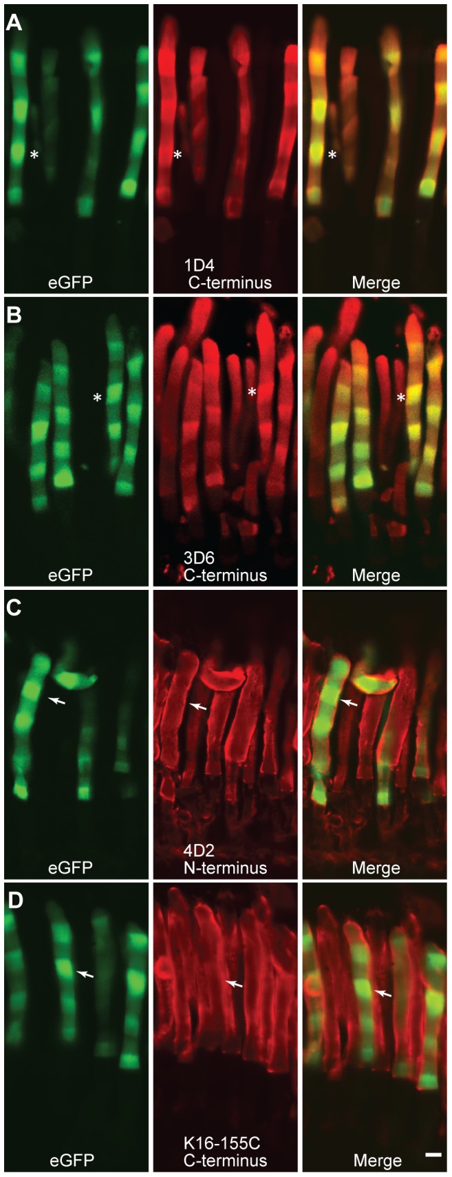
Adult transgenic frogs expressing Rho-eGFP, which has been modified to contain an epitope for the monoclonal antibody 1D4, were kept for 8 weeks in a 168 h (84L–84D) light cycle and then eyes were fixed and immunostained with the indicated antibodies. Rho-eGFP signal (green) is the intrinsic from eGFP fluorescence. The antibodies were detected using a secondary antibody labeled with Cy3 (red). Confocal microscope images of individual and merged channels are presented. Examples of corresponding regions with high Rho-eGFP fluorescence are indicated in A and B by asterisks and in C and D by arrows. White bar is 5 µm.