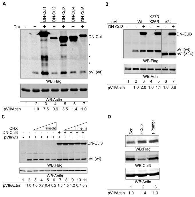
Figure 4. Cullin-3 E3 ubiquitin ligase reduces the Ad5 precursor pVII protein levels.
(A) DN-Cul3 enhances the pVII(wt) protein stability. Expression of the pVII(wt)Flag protein was detected in Dox-induced HEK293-pVII(wt)Flag stable cell line in the presence of transiently overexpressed DN-Cul1-5 plasmids. The pVII(wt) and DN-Cul1-5 proteins contain Flag-tag, which was used to detect the respective proteins by Western blotting. Asterisks (*) indicate degradation products of the DN-Cul proteins. Relative accumulation of the pVII(wt)Flag proteins is shown as the fluorescence signal ratio of pVII/Actin proteins. Hyphen (-) indicates non-Dox induced cells. (B) The pVII(K26R/K27R) and pVII(Δ24) protein levels are not changed by the DN-Cul3 protein overexpression. Expression of the pVII(wt)Flag, pVII(K26R/K27R)Flag, pVII(Δ24)Flag and DN-Cul3-Flag proteins were detected with Flag antibody in HEK293T cells. The ratio of the Flag-tagged pVII proteins to Actin signals (pVII/Actin) is shown after quantification of the fluorescence signals on the Western blot image. Lanes 6 and 7 are derived from the same Western blot image as lanes 1-5. Hyphen (-) indicates non-transfected cells. (C) DN-Cul3 blocks the pVII(wt)Flag protein decay. HeLa cells transiently expressing pVII(wt)Flag in the presence or absence of DN-Cul3-Flag (lanes 7-11) were treated with CHX for 3 hours (lanes 3 and 8), 6 hours (lanes 4 and 9), 9 hours (lanes 5 and 10) and 12 hours (lanes 6 and 11). The expressed proteins were detected with anti-Flag and anti-Actin antibodies. Relative accumulation of the pVII(wt)Flag protein is shown as the fluorescence signal ratio of pVII/Actin proteins. Hyphen (-) indicates non-transfected cells. (D) siRNA knockdown of Cul3 and Psmb1 in HEK293-pVII(wt)Flag cells results in an increase of the pVII(wt)Flag protein levels. After 36 hours of siRNA transfection the pVII(wt)Flag expression was induced with Dox for additional 14 hours. The expression of the proteins was detected by Western blotting with anti-Flag, anti-Cul3 and anti-Actin antibodies. Relative accumulation of the pVII(wt)Flag protein is shown as the fluorescence signal ratio of pVII/Actin proteins.

