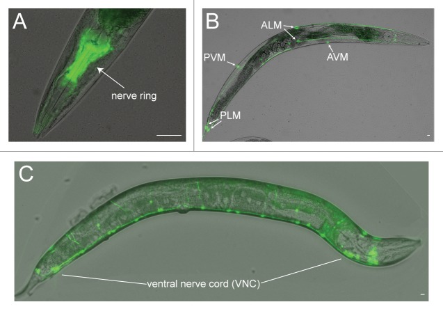
Figure 1. Fluorescence reporter lines enable the visualization of neurons in C. elegans. (A) Nerve ring, positioned between the anterior and terminal bulbs of the pharynx. The sIs11686(Pptl-1::gfp) reporter line is shown.54 (B) Touch receptor neurons, where cell bodies for the AVM, ALMs, PVM and PLMs are shown. The zdIs5(Pmec-4::gfp) reporter line is shown.55 (C) Ventral nerve cord GABAergic motor neurons. The oxIs12(Punc-47::gfp) reporter line is shown.56 Scale = 10 µm.

An official website of the United States government
Here's how you know
Official websites use .gov
A
.gov website belongs to an official
government organization in the United States.
Secure .gov websites use HTTPS
A lock (
) or https:// means you've safely
connected to the .gov website. Share sensitive
information only on official, secure websites.
