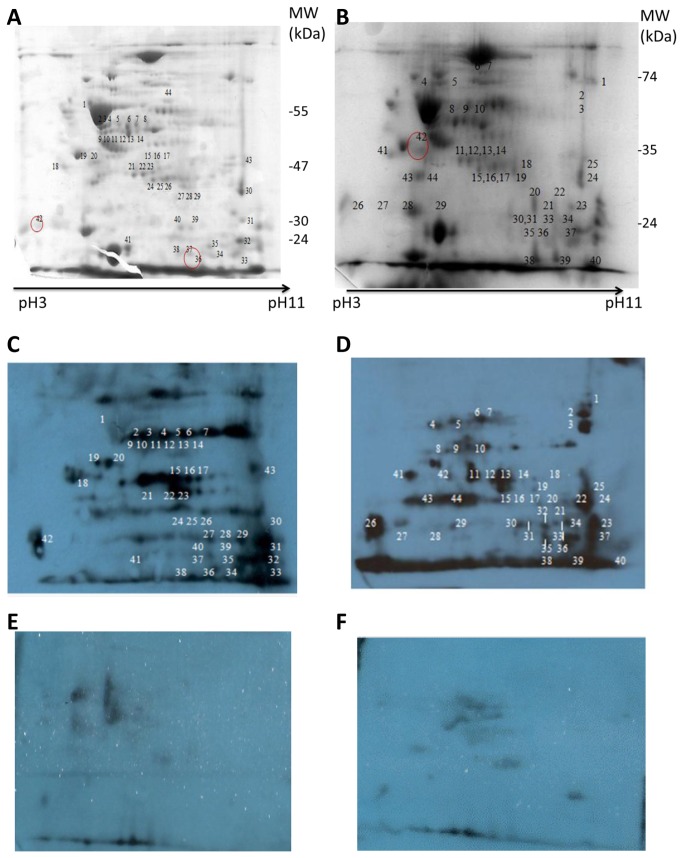
Figure 3. Representative Coomassie blue stained 2-D gel (pI 3-11) of B. multivorans strains, LMG13010 (A, E) and C1962 (B) and the corresponding Western blots probed with pooled patient serum from Bcc colonised patients (C and D) or with serum from patients with no history of Bcc colonisation (E and F).
Each 12 % 2-D gel was prepared with membrane protein preparations extracted from 18 h cultures grown at 37° , and focused on IEF strips (pH 3 to pH 11), blotted and probed the respective sera and detected with anti-human IgG. The corresponding spots on the gel was excised and identified by MALDI-ToF/MS analysis. The numbers represent the proteins spots referred to in the text. The images shown are representative of three individual experiments.

