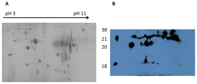
Figure 4. Representative Coomassie blue stained 15% gels and corresponding blots for B. multivorans strain LMG13010.
Each gel was prepared with 120 µg membrane protein preparations extracted from 18 h stationary phase cultures, focussed on pH gradient strips (pH 3 to pH 11), separated on 15% SDS PAGE gels and either stained with Coomassie blue (A) or probed with serum from Bcc colonised CF patients (B). The approximate positions of molecular mass markers (kDa) are indicated beside the Coomassie blue stained gel.

