
Figure 2. Photoreceptor differences between pigmented and albino deer mice.
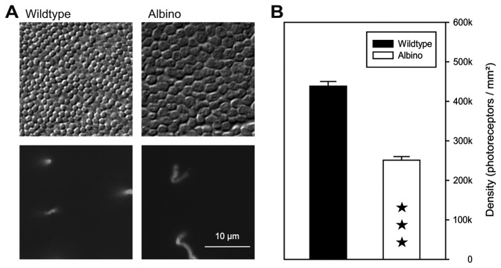
(A) Top: flat view of photoreceptor inner segments in wildtype and albino animals, showing the larger IS diameter and lower density of the albino photoreceptors, differential interference contrast images. Bottom: cones in the same fields, combined immunofluorescence labeling for M and S opsin. The scale bar applies to all images. (B) Quantification of the photoreceptor densities in wildtype and albino. Counts were made at several positions across the retina (wildtype: 11 positions in 1 retina; albino: 6 positions in 2 retinae of 2 individuals; data given as mean and SEM. ★★★, difference statistically significant at p<0,001 (t-test).