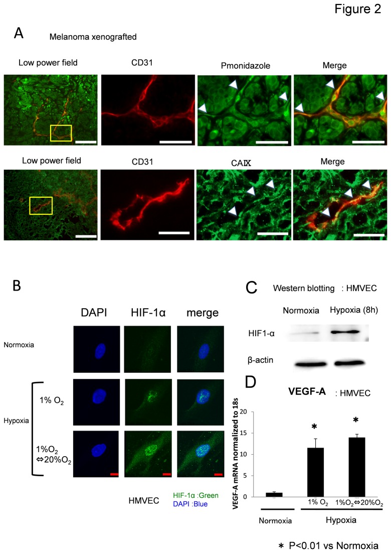
Figure 2. Hypoxia induces HIF-1α and VEGF-A expression in ECs.
(A) The hypoxic area in human tumor xenografts in nude mice was analyzed using the hypoxia marker pimonidazole and CA IX. Tumor tissues were double-stained with anti-CD31 (red) and anti-pimonidazole antibodies or anti-CA IX (green) to visualize hypoxic areas. Pimonidazole staining revealed that tumor vessels were exposed to hypoxia to some extent. Scale bar, 100 μm. (B) HMVECs were cultured and treated for 8 h under normoxia or hypoxia. HIF-1α protein was upregulated 8 h after hypoxia, as revealed by western blotting. Densitometry analysis revealed that HIF-1α was induced by hypoxia. (C) HMVECs were cultured and treated for 8 h under normoxia or hypoxia. HIF-1α protein was upregulated 8 h after hypoxia, as revealed by western blotting. Densitometry analysis revealed that HIF-1α was induced by hypoxia. The experiment was repeated three times. Representative data is shown. (D) mRNA levels of VEGF-A were significantly increased by hypoxia in HMVECs. Experiments were performed in triplicate. *p < 0.01.

