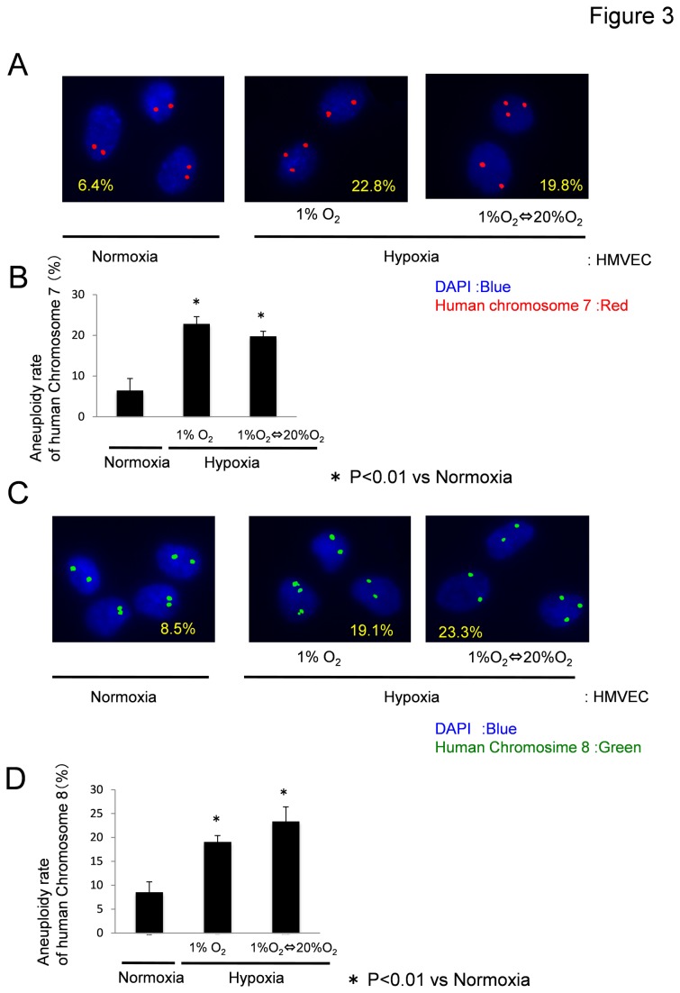
Figure 3. Hypoxia induces aneuploidy in HMVECs.
(A, B) After HMVECs were cultured in each condition for 7 days, FISH analysis using a chromosome-7 probe revealed that approximately 22.8% ECs in the hypoxic condition (1% O2) and 19.8% ECs in the hypoxia-reoxygenation condition were aneuploid, whereas 6.4% ECs in the normoxic condition were aneuploid. (C, D) FISH analysis using a chromosome-8 probe revealed that approximately 19.1% ECs in the hypoxic condition and 23.3% of ECs in the hypoxia-reoxygenation condition were aneuploid, whereas 8.5% ECs in the normoxic condition were aneuploid.

