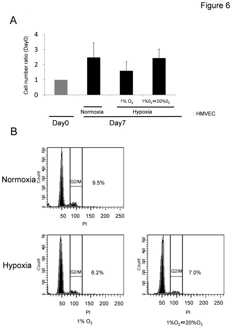
Figure 6. Association between cell cycle and EC aneuploidy in HMVECs exposed to hypoxia.
(A) Cell number and the cell cycle in HMVECs exposed to hypoxia were analyzed by flow cytometry after staining fixed cells with propidium iodide. In each phase, cultured ECs proliferated for 7 days (normoxia, 2.5-fold; hypoxia (1% O2), 1.6-fold; hypoxia-reoxygenation, 2.4-fold versus Day 0 cell numbers). (B) We analyzed the distribution of ECs throughout the cell cycle by flow cytometry. There was no significant difference in cell-cycle distribution between conditions of normoxia and hypoxia. The experiment was repeated three times, and representative data is shown.

