
Fig. 1.
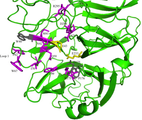
N-terminal part of the crystal structure of endo-inulinase INU2 from A. ficuum with the mutated residues indicated and coloured in purple. The two catalytic residues, E43 and E 233, are shown in yellow. The conserved W40, D175 and P176 are shown in grey. (For interpretation of the references to colour in this figure legend, the reader is referred to the web version of this article.)