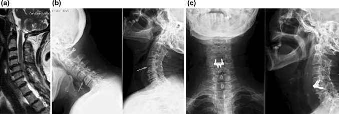
Fig. 1.

a Sagittal, T2-weighted MRI scan showing degenerative disc disease and spinal cord compression at C4–C5 and C5–C6; signs of instability at C4–C5 are also seen. b Lateral flexion–extension X-ray confirming instability at C4–C5. c Postoperative X-ray showing a hybrid construct with a Zero-P device at C4–C5 (the unstable level) and a stand-alone CFRP cage at C–C6 level
