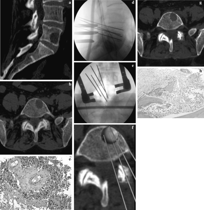
Fig. 2.
M.A. 61 years, L5 melanoma metastases (a, b). A histological slide obtained by a CT-trocar biopsy confirmed the diagnosis (tissue positive to MART 1) (c). After an open laminectomy, using navigator system, four needles were positioned around the lesion and electroporation was performed (d, e, f). A good local control of the disease was confirmed by a CT-trocar biopsy performed 6 months after surgery (necrotic tissue) (g, h)

