
Fig. 1.
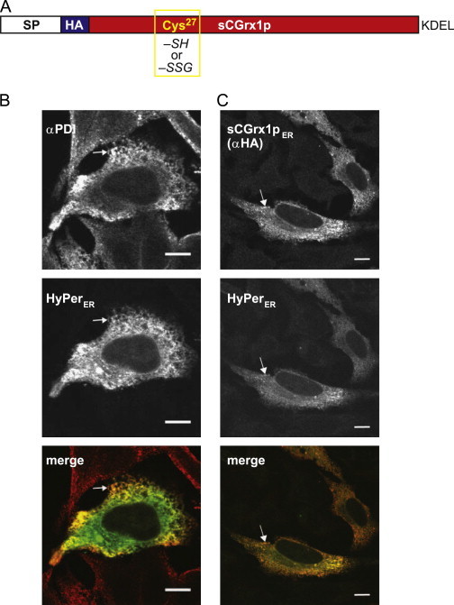
Targeting of sCGrx1p to the ER. (A) Schematic representation of sCGrx1pER. The active site Cys27 is either reduced (–SH) or glutathionylated (–SSG). SP, ER signal peptide; HA, hemagglutinin epitope; KDEL, ER retrieval motif. (B) HyPerER (green) was transfected into HeLa cells and the cells stained with αPDI followed by a red-fluorescent goat-anti-mouse antibody. (C) HeLa cells were co-transfected with HyPerER (green) and sCGrx1pER, which was stained with αHA/goat-anti-mouse (red). Merged images are shown in the bottom panel, and white arrows highlight examples of co-localizing structures. (For interpretation of the references to color in this figure legend, the reader is referred to the web version of this article.)