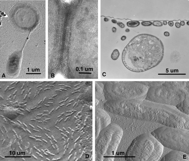
FIG. 4.
TEM, SEM, and AFM of AWI collections. (A) A platinum-shadowed preparation showing a bacterium suspended from a halo-like flotation appendage. (B) A uranyl acetate-negative stain of s-particles at the interface between two prokaryote cells from a tightly packed, raft-like microcolony. (C) The collodion membrane layer with its attached AWI organisms was removed from the slide, fixed, embedded, and sectioned for TEM. The continuous horizontal line is the membrane with attached bacteria, and the large circular object is a section through a protozoan with at least one flagellum. (D) Air-dried SEM sample shows relief image of prokaryotes and various unknown patches in this view of the AWI as seen from beneath the water surface. (E) Atomic force micrograph of the same sample seen in the previous panel.

