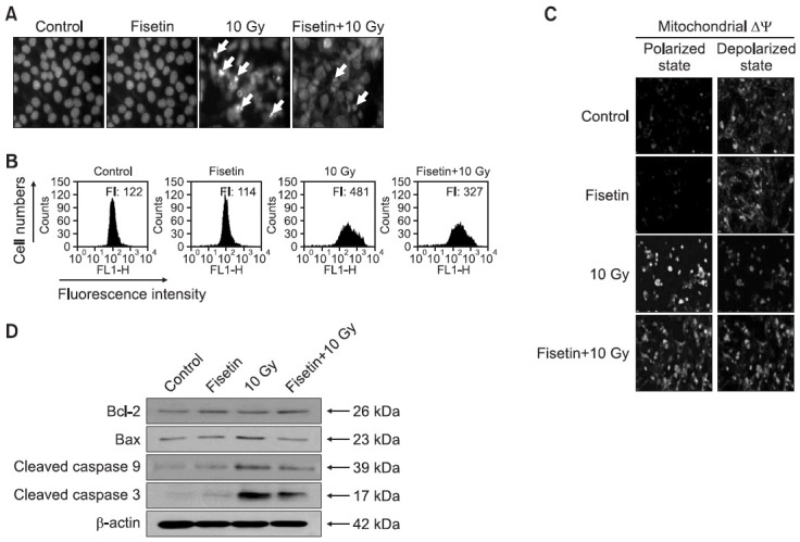
Fig. 4. Mechanisms by which fisetin protects against γ-irradiation-induced apoptotic cell death. Cells were treated 2.5 μg/ml fisetin and 1 h later were exposed to 10 Gy of γ-irradiation, and then incubated for 48 h. (A) Formation of apoptotic bodies (arrows) was examined in cells stained with Hoechst 33342. (B, C) The mitochondrial membrane potential (Δψm) of JC-1-stained cells was analyzed by flow cytometry and confocal microscopy. (D) Cell lysates were electrophoresed and Bcl-2, Bax, cleaved caspase-9, and cleaved caspase-3 were detected by immunoblot analysis using the corresponding.

