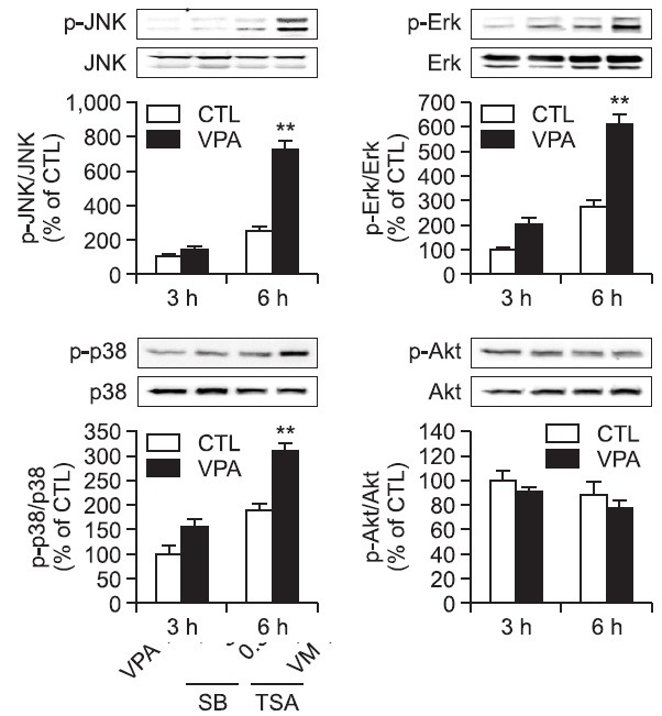
Fig. 4. The activation of MAPK pathways by VPA in rat primary astrocytes. Rat primary astrocytes in serum-free DMEM/F12 were treated with VPA (1 mM) at 37℃. Cell extracts were collected for Western blot analysis. Immunoblots of lysates form treated rat primary astrocytes were probed with phospho-ERK, phospho-p38, phospho-JNK, and phosphop-Akt antibodies. As a loading control, total ERK/JNK/p38/Akt levels were also measured. **p<0.01 vs. control.

