Abstract
We present a case of a 46-year-old man with a history significant only for hypertension and depression that presented with a new onset seizure resulting from a right parietal lobe mass. Further evaluation determined the parietal mass to be central nervous system toxoplasmosis, which was the initial presentation of his underlying HIV/AIDS. This case provided a diagnostic challenge and demonstrates the importance of a thorough evaluation as it pertains to a newly diagnosed brain lesion.
Background
Seizures are a relatively common initial manifestation of a space-occupying brain lesion. The differential diagnoses of a new onset seizure are vast and therefore it is essential for the clinician to perform a thorough evaluation. CT and MRI are often the initial diagnostic tests used in the setting of a new onset seizure to evaluate for any space occupying lesions. If present, the characteristics of the lesion seen on imaging may be suggestive of a neoplastic or infectious aetiology, however as this case demonstrates, should not preclude further exploration of other causes.
Case presentation
A 46-year-old man, with a medical history significant only for hypertension and depression, presented to an outside hospital with a witnessed episode of a new onset seizure. As per family accounts, he was performing yard work for the greater portion of the day in extreme heat after which he came home and sat down at the dinner table of hunger and dehydration when the episode ensued. The family described the episode as echolalia followed by 3–5 min of generalised tonic–clonic activity. Once the seizure had subsided, the patient was unaware of his surroundings and unable to recognise his family members for 20–30 min. Emergency medical services were called and the patient arrived to the emergency department (ED) through ambulance no longer in a postictal state.
Differential diagnosis
Primary brain neoplasm (glioblastoma multiforme)
Central nervous system (CNS) lymphoma
Bacterial or fungal abscess
CNS toxoplasmosis
Treatment
At the outside hospital, an initial CT scan was performed which revealed a white matter low-density change in the right deep high parietal lobe. At this point the patient was transferred to a tertiary care facility for further evaluation and neurosurgical intervention.
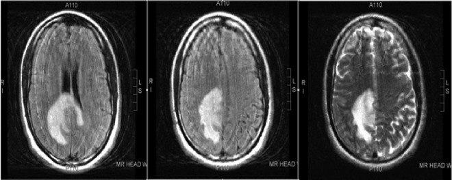
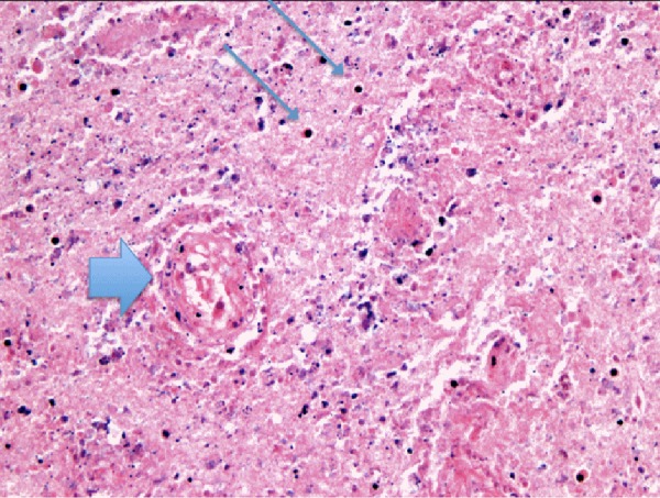
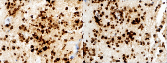
Once transferred to the tertiary care facility the patient was given intravenous dexamethasone and a loading dose of phenytoin. MRI was performed to further characterise the lesion. The results of MRI confirmed the findings on CT describing the lesion as a ring-enhancing mass in the deep right parietal lobe which was most concerning for a primary brain neoplasm such as glioblastoma multiforme or less likely lymphoma (figure 1). Neurosurgery then performed a biopsy demonstrating significant necrosis, which can be consistent with glioblastoma multiforme. Interestingly, however, there was no evidence of malignant cells seen in the limited viable tissue of the biopsy specimen which raised concern for the possibility of an infectious aetiology be it bacterial or fungal in origin (figure 2). The patient was then tested for HIV and was found to be positive on initial antibody screening with subsequent positive confirmatory western blot. His CD4 count was measured to be 17 and as a result was further evaluated for any opportunistic infections, namely toxoplasmosis as the culprit. Serum toxoplasma IgG was positive at greater than 900 IU/mL (≥12 IU/mL is positive indicating IgG antibody to Toxoplasma gondii detected which may indicate current or past T gondii infection). Subsequently, the patient's biopsy sample was sent for toxoplasmosis-specific staining which revealed the aetiology of his brain lesion (figure 3). The patient was started on leucovorin, sulfadiazine and pyrimethamine with plans to start highly active antiretroviral therapy (HAART) as an outpatient.
Figure 1.

MRI demonstrating ring-enhancing mass in the right parietal lobe.
Figure 2.

Brain biopsy specimen demonstrating a blood vessel with coagulative necrosis (large arrow) and nuclear debris/necrosis (thin arrows). The overall image is without definition or detail due to extensive necrosis.
Figure 3.

Brain biopsy with immunoperoxidase stains (brown is the positive ‘horseradish peroxidase’ reaction) against antigens for Toxoplasma species. The larger stained objects are cysts and the smaller, slightly oval ones are tachyzoites of Toxoplasma.
Outcome and follow-up
The patient improved during the course of hospitalisation. His hospital course was complicated by several episodes of auditory, visual and olfactory hallucinations. He claimed to have heard the voices of and seen family members not present in addition to recurrent phantosmia, which the patient described as that of a dead rodent. His hallucinations initially attributed to steroid psychosis quickly subsided once his dexamethasone was tapered to discontinuation. He made a full recovery and is currently being followed as an outpatient with plans to start HAART once genotyping results become available.
Discussion
The advent of HAART has significantly reduced the morbidity and mortality associated with HIV. As a result, this disease once thought of as a ‘death sentence’ is now considered to be a chronic manageable condition. Despite advances in therapy, however, patients are consistently being diagnosed in the latter stages of the disease process. Several reports have estimated that 30–40% of persons with HIV have already reached the point of AIDS (as defined by a CD4 count of <200) at the time of diagnosis.1 Early detection is of utmost importance as studies have shown a direct correlation with improved prognosis in patients’ initiating HAART at a higher CD4 count.2
The US Preventative Service Task Force (USPSTF) recently updated its 2005 recommendations on HIV screening guidelines. The updated guidelines recommend clinicians to screen adolescents and adults from the ages of 15–65 for HIV infection. Patients younger than 15 years should be screened if risk factors for HIV transmission are present and similarly patients older than 65 years should be screened if there are on-going risk factors for HIV transmission. The USPSTF states those who are active intravenous drug misusers, have unprotected vaginal or anal intercourse, multiple sexual partners, sexual partners who are positive for HIV and homosexual males as highly at risk for acquiring HIV infection.3
Toxoplasmosis, an infection caused by the intracellular protozoan parasite T gondii, is the most common opportunistic infection of the CNS in patients with AIDS. Initially latent, the parasite reactivates and causes infection when the patients CD4 count falls below 100 cells/mm3, as was the case in our patient.4 Patients with toxoplasmic encephalitis typically present with headache, as did our patient stating he had been experiencing worsening nocturnal headaches for 1 month proceeding the seizure. In one retrospective analysis of 115 patients with toxoplasmosis, 55%, 52% and 47% of patients presented with headache, confusion and fever, respectively.5
The diagnosis of toxoplasmosis is primarily through imaging and brain biopsy. Serology can be obtained however it is not particularly helpful as toxoplasma IgG are positive in the vast majority of patients and IgM antibodies are typically absent even in the acute setting.6 MRI and CT imaging tend to show multiple ring-enhancing brain lesions with predilection towards the basal ganglia.7 Levy et al performed a prospective study on 50 neurologically symptomatic AIDS patients. Each patient was evaluated using both modalities; MRI was better than CT for the detection of intracranial pathology and significantly altered the diagnosis and treatment in 40% of these patients.8 Thallium single photon emission CT (SPECT) and positron emission tomography (PET) have been shown as useful adjunctive imaging to further delineate toxoplasmosis or other infection from CNS lymphoma as it has increased thallium uptake on SPECT and greater glucose and methionine metabolism on PET.9
Learning points.
The characteristics of space occupying lesions seen on CT or MRI may be suggestive of a neoplastic or infectious aetiology; however, this should not preclude further exploration of other causes.
Toxoplasmosis is a protozoan parasite which reactivates and causes infection when the patients CD4 count falls below 100 cells/mm.
MRI or CT imaging with multiple ring-enhancing brain lesions with predilection towards the basal ganglia may be suggestive of toxoplasmosis.
MRI because of its great sensitivity might be the best neuroimaging modality for the evaluation of neurologically symptomatic patients with AIDS.
Single photon emission CT or positron emission tomography may be useful in differentiating toxoplasmosis and other infection from central nervous system lymphoma.
The treatment of choice for toxoplasmosis is pyrimethamine, sulfadiazine and leucovorin for a 6-week duration. Alternative regiments for the treatment of toxoplasmosis include pyrimethamine plus clindamycin, pyrimethamine plus azithromycin, pyrimethamine plus atovaquone or trimethoprim–sulfamethoxazole.
The US Preventative Service Task Force (USPSTF) recently updated its HIV screening guidelines, recommending that clinicians should screen all patients from 15 to 65 years of age for HIV as well as all pregnant women including those who are present in labour and are untested, and those whose HIV status is unknown. If risk factors are present, earlier screening or continued screening is appropriate. Further studies need to be performed to determine optimum intervals for HIV screening.
Footnotes
Contributors: All authors have contributed to this case report, have reviewed the final version and approve it for publication.
Competing interests: None.
Patient consent: Obtained.
Provenance and peer review: Not commissioned; externally peer reviewed.
References
- 1.Klein D, Hurley LB, Merrill D, et al. Review of medical encounters in the 5 years before a diagnosis of HIV-1 infection: implications for early detection. J Acquir Immune Defic Syndr 2003;2013:143–52 [DOI] [PubMed] [Google Scholar]
- 2.Kaplan J, Hanson D, Karon J, et al. Late initiation of antiretroviral therapy (at CD4+ lymphocyte count <200 cells/mL) is associated with increased risk of death [abstract 520]. 8th Conference on Retroviruses and Opportunistic Infections, February 4–8, 2001, Chicago, IL, USA [Google Scholar]
- 3. http://annals.org/article.aspx?articleid=1682314 (accessed 16 Jun 2013) [Google Scholar]
- 4.Renold C, Sugar A, Chave JP, et al. Toxoplasma encephalitis in patients with the acquired immunodeficiency syndrome. Medicine (Baltimore) 1992;2013:224–39 [DOI] [PubMed] [Google Scholar]
- 5.Porter SB, Sande MA. Toxoplasmosis of the central nervous system in the acquired immunodeficiency syndrome. N Engl J Med 1992;2013:1643–8 [DOI] [PubMed] [Google Scholar]
- 6.Luft BJ, Brooks RG, Conley FK, et al. Toxoplasmic encephalitis in patients with acquired immune deficiency syndrome. JAMA 1984;2013:913–17 [PubMed] [Google Scholar]
- 7. http://www.uptodate.com/contents/toxoplasmosis-in-hiv-infected-patients?source=search_result&search=toxoplasmosis&selectedTitle=2~150#H6 (accessed 16 Jun 2013) [Google Scholar]
- 8.Levy RM, Mills CM, Posin JP, et al. The efficacy and clinical impact of brain imaging in neurologically symptomatic AIDS patients: a prospective CT/MRI study. J Acquir Immune Defic Syndr 1990;2013:461–71 [PubMed] [Google Scholar]
- 9.Skiest DJ, Erdman W, Chang WE, et al. SPECT thallium-201 combined with Toxoplasma serology for the presumptive diagnosis of focal central nervous system mass lesions in patients with AIDS. J Infect 2000;2013:274–81 [DOI] [PubMed] [Google Scholar]


