Figure 2.

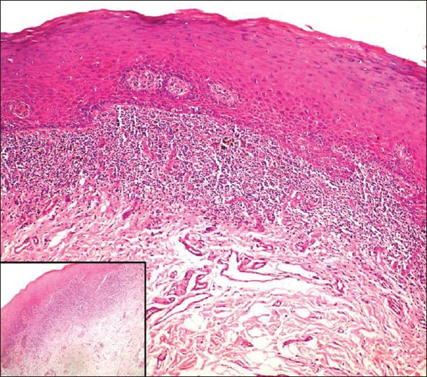
Atrophic lichen planus; showing a dense juxtaepithelial lymphocytic infiltrate (H & E stain, ×100); Inset: Higher magnification (H & E stain ×400)

Atrophic lichen planus; showing a dense juxtaepithelial lymphocytic infiltrate (H & E stain, ×100); Inset: Higher magnification (H & E stain ×400)