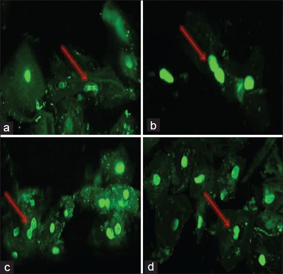
Figure 2.

Patients with clinically diagnosed and/or suspected squamous cell carcinoma -.cases. (a-d) Acridine orange confocal microscopy, ×400, showing numerous abnormal mitotic figures at different stages of mitosis. a shows anaphase, b shows starting of telophase, c shows middle of telophase, and d shows ending of telophase of mitosis compound microscope, ×100 and ×400, respectively, showing abnormal mitotic figures
