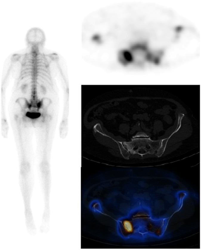
Figure 2.
Patient with a 2-year history of breast cancer, a background of degenerative spine disease and left total-hip replacement presenting with pain in the right sacroiliac area failing to settle 4 months following a fall. On the planar images, there is intense tracer uptake seen in both sacroiliac regions, right more than left. Low-grade tracer uptake is seen in both shoulders; left tenth rib posteriorly and lower lumbar spine. Note is made of left hip prosthesis. On the single photon emission CT-CT, the avid tracer uptake is localised to the right sacral ala. The right side of the sacrum shows more sclerosis than the left side with vertical linear lucency. Scan appearances in keeping with a right sacral ala fracture.

