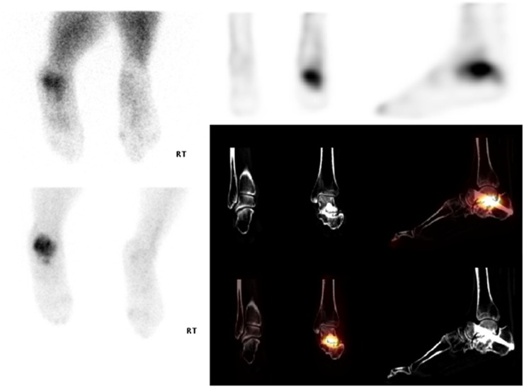
Figure 7.
Patient with left subtalar joint fusion but persistent pain. On the delayed views of the feet (top right and bottom left), there is increased tracer uptake seen involving the left ankle. On the early blood pool images (top left), there is increased vascularity seen to this site. On the single photon emission CT-CT images within the left foot, the increased area of uptake corresponds to the intended subtalar fusion. There is persistent joint space noted with significant degenerative changes. The appearances are in keeping with non-union of the intended subtalar fusion.

