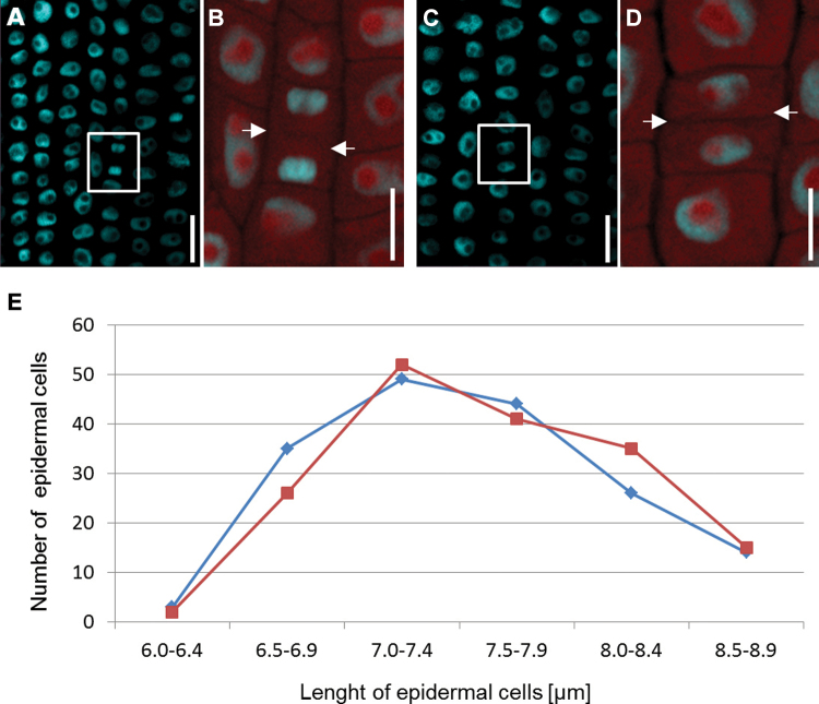
Fig. 5.
Shootward-last division of epidermal cells in a cv. ‘Karat’ root. (A) The highlighted region indicates daughter cells at late mitosis. (B) Fluorescence of the cytoplasm identifies the cell plate (indicated by arrows). (C) Compact nuclei of epidermal cells immediately post-cytokinesis. (D) Cytoplasm fluorescence, permitting an estimate of cell length (arrows indicate the cell wall separating adjacent daughter cells). Nuclei in (A)–(D) were stained with DAPI. Bars, 10 μm. (E) Lengths of the shootward (blue) and rootward (red) cells after the shootward-last cell division.

