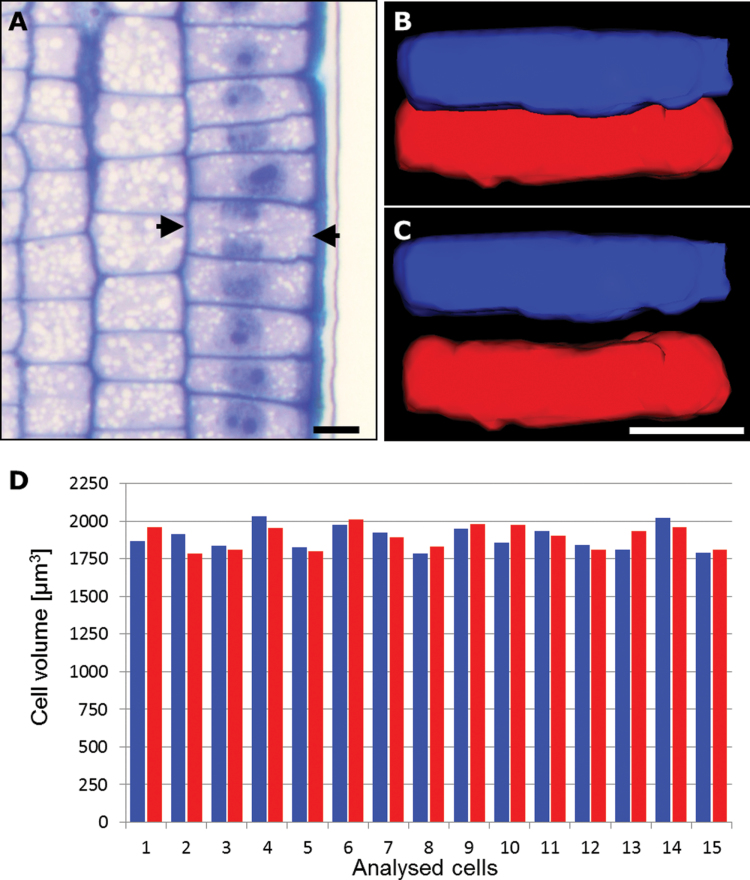
Fig. 6.
Volume of daughter cells following the shootward-last cell division. (A) Longitudinal sections through a cv. ‘Karat’ root (used for 3D reconstruction). Arrows indicate the cell plate separating adjacent daughter cells. (B, C) 3D modelling of shootward (blue) and rootward (red) cells: in vivo arrangement (B) and isolated cells (C). Bars, 20 μm. (D) Cell volumes of shootward (blue) and rootward (red) cells.

