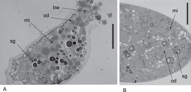
Fig. 5.
Transmission electron microscopic sections of Lilium pollen tubes. (A) Bursting tube near the tip showing ejection of cell contents after Hg. The organelles are intact and similar to those of growing tubes. (B) Growing tube without Hg showing cell contents. Bw, bursting wall; od, oil droplet; mi, mitochondrion; sg, starch granule. Bars=5 µm.

