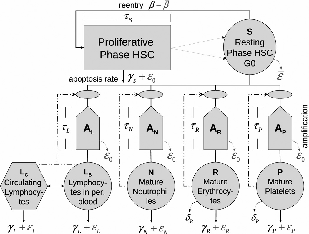
Fig 1.

Model of hematopoiesis under chronic exposure to IR. The circles describe five model compartments corresponding to HSC (S) and mature lymphocytes (L), neutrophiles (N), erythrocytes (R), and platelets (P). The pentagons denote compartments for amplification and maturation of blood cells. The rectangle shows the compartment of HSC proliferation. The hexagon shows the compartment of circulating lymphocytes. Solid lines show cell transitions between compartments, while dash-dotted lines show feedback loops regulating the peripheral blood counts. The solid lines with arrows represent different modes of cell death: i)γ’s correspond to natural apoptosis, ii)ε’s correspond to radiation-induced apoptosis, and iii)δ’s correspond to senescence death of platelets and erythrocytes.
