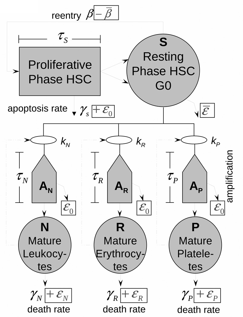
Fig. 1.
The model of hematopoiesis under chronic exposure to IR. The circles describe four model compartments corresponding to HSC (S) and mature leukocytes (N), erythrocytes (R), and platelets (P). The pentagons denote compartments for amplification and maturation of blood cells. The square shows the compartment of HSC proliferation. Solid lines show the cell transitions between compartments, and dash-dotted lines show feedback loops regulating the blood counts.

