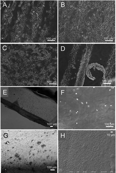
Figure 7.

Representative optical images of adherent NIH3T3 fibroblasts on PS substrates coated with PNIPAM micropatterns of 100 μm stripes/100 μm spacings at 37 °C for 24 hr (A), 48 hr (B) and 72 hr (C), and detached fibroblasts upon lowering the temperature to 25 °C (D, E). On the substrate after cell detachment (F), the PNIPAM particles were observed under DIC microscope (G) and SEM (H).
