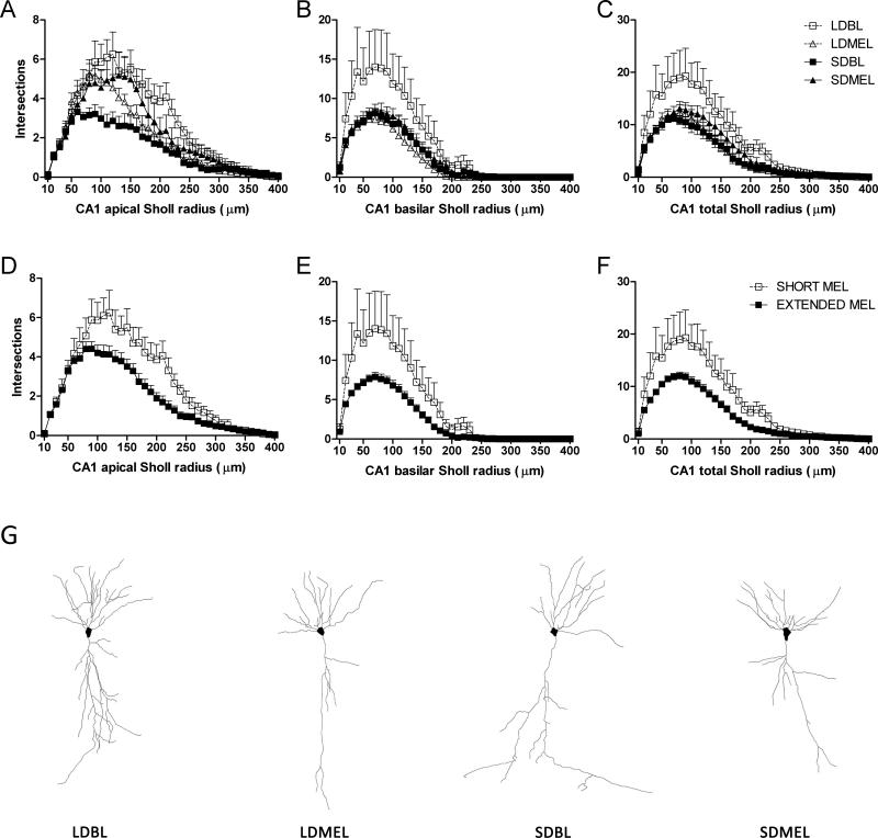
Figure 5.
CA1 dendritic complexity. Extended duration of melatonin exposure reduces complexity in CA1 apical (A,D), basilar (B,E), and total (C,F) dendritic fields. Representative Neurolucida tracings of CA1 pyramidal neurons from each experimental group (G). Data in D, E, and F are collapsed by melatonin exposure duration from A, B, and C respectively. A, B, and C share key depicted in C. D, E, and F share key depicted in F.

