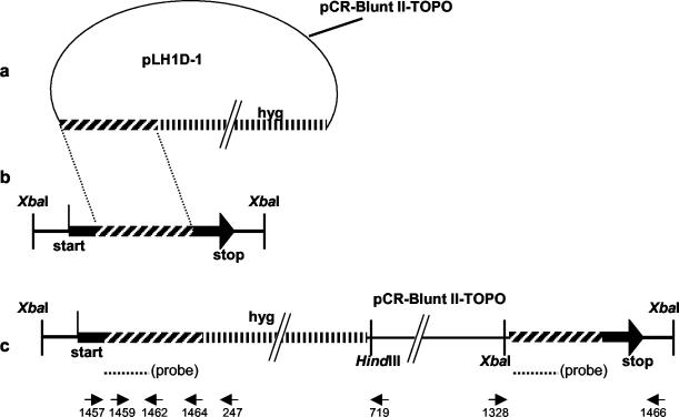
FIG. 1.
Disruption of LH1 in F. graminearum. (a) Diagram of disruptant plasmid pLH1D-1. The hatched segment on the left represents a 1,281-bp fragment of LH1. The vertically lined segment on the right represents a promoter 1-hygB fragment. (b) Diagram of the Z-3639 genomic DNA containing the LH1 region. “Start” and “stop” are the ends of LH1. (c) Diagram of the LH1 region of a transformant resulting from a single crossover event. The binding position of the probe used for Southern analysis is indicated; arrows indicate the positions of primers. Primer sequences are given in Table 1.

