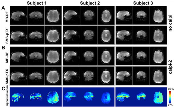
FIG. 6.

Three-plane view of the whole-brain EPI acquisitions with axial slices in three subjects, acquired at factor-2 slice acceleration with MB-RF and SMS-pTX excitations. Data acquired without FOV-shift between simultaneous slices are shown in panel (A); repeating the same experiment with blipped-CAIPIRINHA (FOV/2-shift between slices; panel (B)) did not visibly affect image quality, which demonstrates the suitability of the dual-row geometry to separate two axial slices. Overall apparent image quality between the MB-RF and SMS-pTX methods is comparable, but signal reductions and discontinuities in image intensity can be observed in the transition areas between the two slice groups. This is attributable to the reduced B1+ especially near interface of the two coil rows and also the step function in B1+ profile, as is evident from the maps in panel (C), which show the relative signal reduction of SMS-pTX vs. MB-RF (for the case without CAIPI).
